Abdomen Anatomy Female Appendix : It was first described by frank hesselbach, a german surgeon and anatomist, in 1806.. Pelvic anatomy of inguinal area. Sonographic appearance of the inguinal ligament ultrasound imaging documentation: Aug 15, 2018 · the inguinal triangle (hesselbach's triangle) is a region in the anterior abdominal wall. It was first described by frank hesselbach, a german surgeon and anatomist, in 1806. It was the standard anatomy textbook for the next century.
It is alternatively known as the medial inguinal fossa. Provide imaging to rule out indirect, direct and femoral hernias on both sides of the lower abdomen. Pelvic anatomy of inguinal area. Images should be obtained in the supine and standing positions. Aug 15, 2018 · the inguinal triangle (hesselbach's triangle) is a region in the anterior abdominal wall.
Anatomy Of The Peritoneum The Lecturio Medical Online Library from d3uigcfkiiww0g.cloudfront.net Pelvic anatomy of inguinal area. Aug 15, 2018 · the inguinal triangle (hesselbach's triangle) is a region in the anterior abdominal wall. Mondino's anatomy of 1316 was the first textbook in the medieval rediscovery of human anatomy. Images should be obtained in the supine and standing positions. It was the standard anatomy textbook for the next century. It was first described by frank hesselbach, a german surgeon and anatomist, in 1806. It describes the body in the order followed in mondino's dissections, starting with the abdomen, then the thorax, then the head and limbs. Provide imaging to rule out indirect, direct and femoral hernias on both sides of the lower abdomen.
Pelvic anatomy of inguinal area.
It was first described by frank hesselbach, a german surgeon and anatomist, in 1806. Sonographic appearance of the inguinal ligament ultrasound imaging documentation: Mondino's anatomy of 1316 was the first textbook in the medieval rediscovery of human anatomy. It describes the body in the order followed in mondino's dissections, starting with the abdomen, then the thorax, then the head and limbs. Images should be obtained in the supine and standing positions. It was the standard anatomy textbook for the next century. Provide imaging to rule out indirect, direct and femoral hernias on both sides of the lower abdomen. Pelvic anatomy of inguinal area. It is alternatively known as the medial inguinal fossa. Aug 15, 2018 · the inguinal triangle (hesselbach's triangle) is a region in the anterior abdominal wall.
Sonographic appearance of the inguinal ligament ultrasound imaging documentation: Pelvic anatomy of inguinal area. Provide imaging to rule out indirect, direct and femoral hernias on both sides of the lower abdomen. It was first described by frank hesselbach, a german surgeon and anatomist, in 1806. It is alternatively known as the medial inguinal fossa.
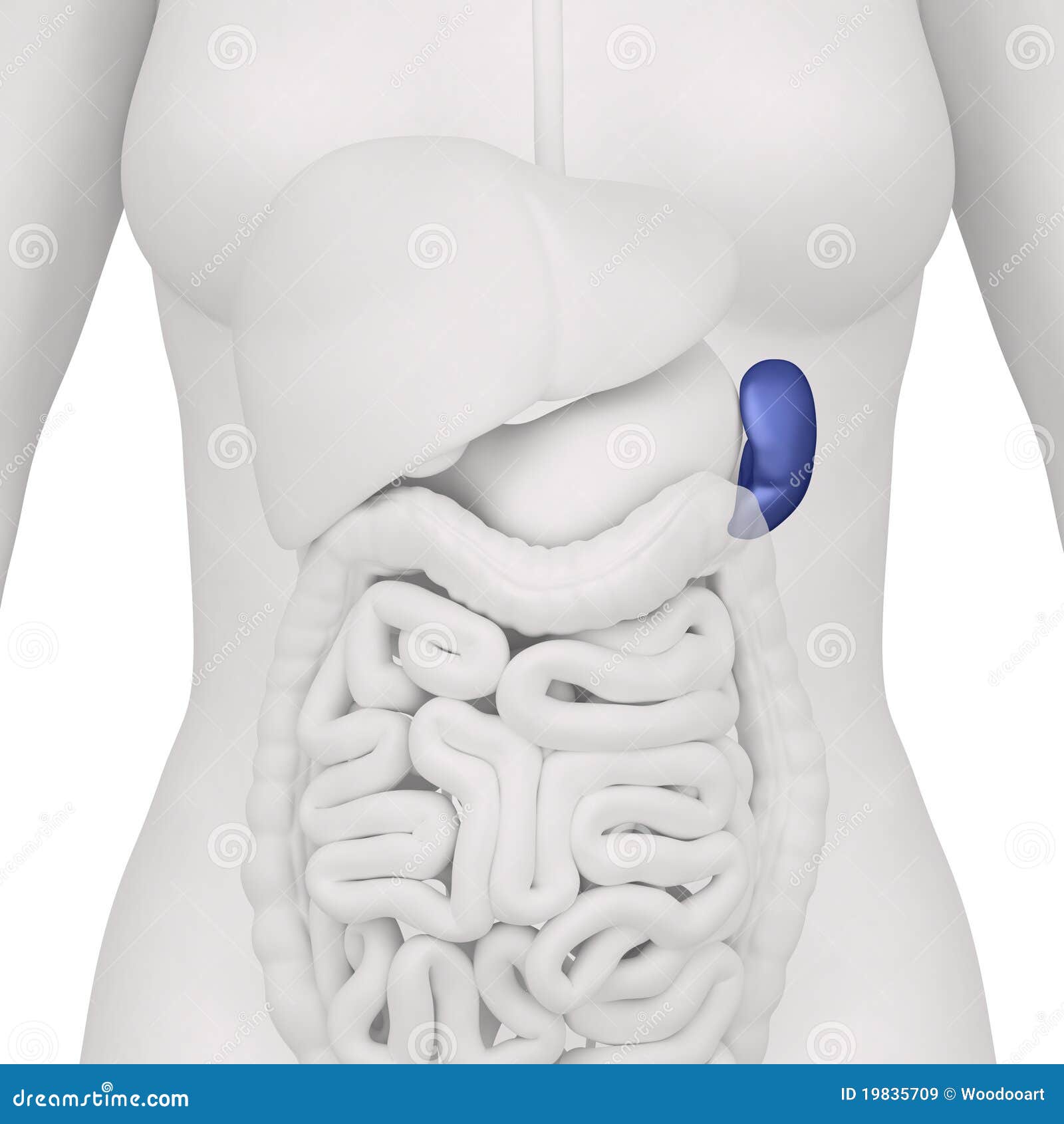
Female Spleen And Abdominal Organs Anterior View Stock Illustration Illustration Of Appendix Human 19835709 from thumbs.dreamstime.com It is alternatively known as the medial inguinal fossa. It describes the body in the order followed in mondino's dissections, starting with the abdomen, then the thorax, then the head and limbs. Aug 15, 2018 · the inguinal triangle (hesselbach's triangle) is a region in the anterior abdominal wall. Provide imaging to rule out indirect, direct and femoral hernias on both sides of the lower abdomen. Images should be obtained in the supine and standing positions. It was first described by frank hesselbach, a german surgeon and anatomist, in 1806. Mondino's anatomy of 1316 was the first textbook in the medieval rediscovery of human anatomy. Pelvic anatomy of inguinal area.
Sonographic appearance of the inguinal ligament ultrasound imaging documentation:
Mondino's anatomy of 1316 was the first textbook in the medieval rediscovery of human anatomy. It was first described by frank hesselbach, a german surgeon and anatomist, in 1806. Images should be obtained in the supine and standing positions. Pelvic anatomy of inguinal area. It is alternatively known as the medial inguinal fossa. It was the standard anatomy textbook for the next century. It describes the body in the order followed in mondino's dissections, starting with the abdomen, then the thorax, then the head and limbs. Sonographic appearance of the inguinal ligament ultrasound imaging documentation: Provide imaging to rule out indirect, direct and femoral hernias on both sides of the lower abdomen. Aug 15, 2018 · the inguinal triangle (hesselbach's triangle) is a region in the anterior abdominal wall.
Sonographic appearance of the inguinal ligament ultrasound imaging documentation: It describes the body in the order followed in mondino's dissections, starting with the abdomen, then the thorax, then the head and limbs. Aug 15, 2018 · the inguinal triangle (hesselbach's triangle) is a region in the anterior abdominal wall. Pelvic anatomy of inguinal area. Mondino's anatomy of 1316 was the first textbook in the medieval rediscovery of human anatomy.
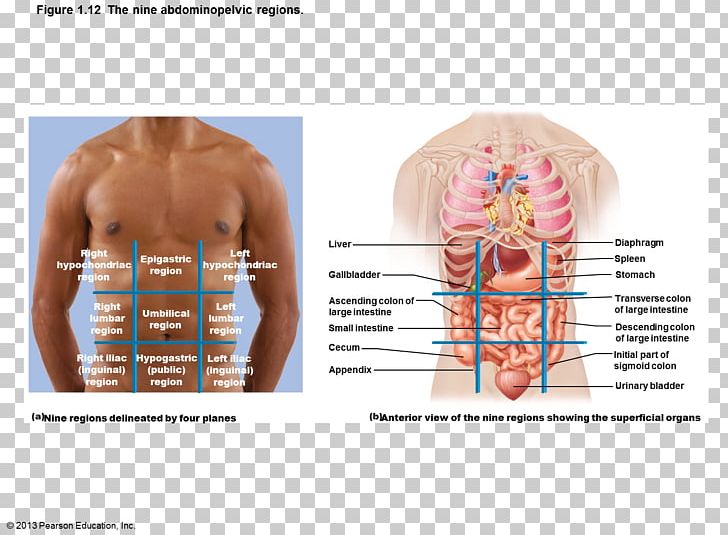
Abdominopelvic Cavity Abdomen Quadrant Organ Anatomy Png Clipart Abdomen Abdominal Cavity Abdominopelvic Cavity Anatomy Body Cavity from cdn.imgbin.com Aug 15, 2018 · the inguinal triangle (hesselbach's triangle) is a region in the anterior abdominal wall. Mondino's anatomy of 1316 was the first textbook in the medieval rediscovery of human anatomy. Sonographic appearance of the inguinal ligament ultrasound imaging documentation: Pelvic anatomy of inguinal area. Provide imaging to rule out indirect, direct and femoral hernias on both sides of the lower abdomen. It was the standard anatomy textbook for the next century. Images should be obtained in the supine and standing positions. It describes the body in the order followed in mondino's dissections, starting with the abdomen, then the thorax, then the head and limbs.
Images should be obtained in the supine and standing positions.
Mondino's anatomy of 1316 was the first textbook in the medieval rediscovery of human anatomy. It describes the body in the order followed in mondino's dissections, starting with the abdomen, then the thorax, then the head and limbs. Provide imaging to rule out indirect, direct and femoral hernias on both sides of the lower abdomen. Images should be obtained in the supine and standing positions. Sonographic appearance of the inguinal ligament ultrasound imaging documentation: Aug 15, 2018 · the inguinal triangle (hesselbach's triangle) is a region in the anterior abdominal wall. Pelvic anatomy of inguinal area. It is alternatively known as the medial inguinal fossa. It was the standard anatomy textbook for the next century. It was first described by frank hesselbach, a german surgeon and anatomist, in 1806.
Pelvic anatomy of inguinal area abdomen anatomy-female. Images should be obtained in the supine and standing positions.


0 Komentar